September case highlights
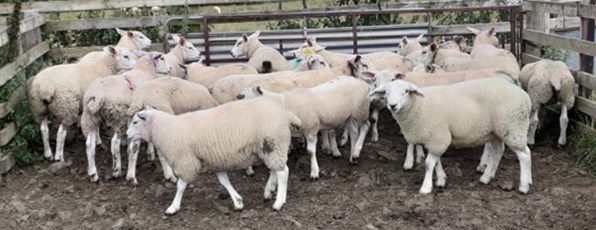
Parasitic disease dominated the diagnoses made in Aberdeen in September with lungworm and Ostertagiasis diagnosed in cattle and gastrointestinal parasites in sheep causing ill thrift and death. Other enteric diseases in cattle included bloat, intestinal torsion, yersiniosis and coccidiosis as well as diet related diagnoses of ruminal acidosis, fog fever and cerebrocortical necrosis. In sheep causes of ill thrift in ewes included Johne’s disease and OPA, while tick borne diseases were also a feature with tick borne fever and louping ill being diagnosed.
Some of the interesting cases finalised in September are featured below:
- Photosensitisation secondary to copper toxicity in sheep
- Fog Fever in a grazing heifer
- Husk and ostertagiasis in a yearling heifer
- Re-infection syndrome in a 20-month-old bull
- Occipital dysplasia leading to sudden death in a Chihuahua puppy
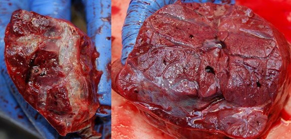
Photosensitisation secondary to chronic copper toxicity. A 6% incidence of photosensitisation in a lowland flock was investigated, with cases noted only in Texel cross lambs. No known plant, fungal or bacterial cause was found at the farm visit. Blood samples were collected from acute and recovering cases, revealing some raised hepatic enzymes especially GGT, and low albumin in chronic cases. Two sheep were submitted for post mortem examination, one in July and a second in late August. Histopathology of the liver in the second case which was a chronically affected animal, was consistent with chronic coper toxicity. Liver and kidney copper assay revealed high levels in both animals. Liver levels were 37,800 and 27,500µmol/kg DM (normal range <7,800µmol/kg DM) and kidney levels were 1850 and 1520 µmol/kg DM (normal range <787µmol/kg DM. Food Safety Scotland was informed. The lambs had been given a 2g copper bolus suitable for animals >25kg in early June, when some lambs may have been as young as 4 weeks old, which resulted in excessive levels of liver copper. There were 5 deaths in early June which were not investigated, and may have been the result of copper toxicity. A subset of animals had liver copper levels which were high enough to result in hepatic dysfunction, leading to photosensitization secondary to hepatic disease. This also led to poor growth rates in the affected lambs. The sensitivity of Texel and Texel cross lambs to high copper intakes explained the breed predisposition within the flock.
Chronic ill thrift and evidence of healing lesions on the ears of affected lambs. One acutely affected animals with oedematous ears seen front right
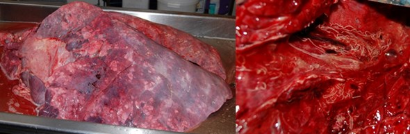
Fog Fever (Acute bovine pulmonary emphysema and edema, ABPEE) in a heifer on silage aftermath. Three weeks after a group of Limousin heifers were moved to a silage aftermath, one animal was noted to be dull, lethargic and breathing heavily for a few days before being found dead. On postmortem examination the lungs were heavy and wet on cut surface, with copious blood-stained fluid within the bronchi. There was an area of bullae formation and interstitial emphysema on the dorsal aspect of the left cranial lung lobes. Histopathologically there was an acute interstitial pneumonia with type II pneumocyte hyperplasia, and in the absence of viral bronchitis or bronchiolitis, fog fever was the most likely cause of the pathology. Histopathology suggested a sub-acute disease course, which is consistent with this case history as fog fever is usually seen within ten days of moving to the lush pasture. Fog fever is an important differential of respiratory disease at this time of year, with clinical respiratory disease often attributed to bacterial pneumonia, or lungworm infection or re-infection syndrome.
Heavy lungs which were wet on cut surface with areas of interstitial bullae formation
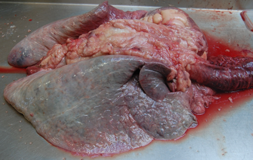
Husk and abomasal parasitism were diagnosed on gross examination of a Simmental-cross yearling heifer submitted to Aberdeen VC. The animal originated from a suckler herd with 60 yearlings in a group, which were wormed and put onto pasture in May. Twelve of the animals in the group showed signs of pneumonia and were unresponsive to antimicrobial treatment. The group had received one dose of Rispoval 4 and an IBR intranasal vaccine. On post-mortem examination, the lungs were heavy and non-collapsed and the tracheobronchial lymph nodes were markedly enlarged. There were patchy areas of consolidation on the cut surface of all lung lobes and live white worms were present in the airways. Pasteurella multocida DNA was detected in the lung by multiplex respiratory PCR and a secondary bacterial infection may have contributed to the lung pathology, albeit characteristic gross changes of bronchopneumonia were not detected. In addition to the lung pathology, the mucosa of the abomasum appeared inflamed, and prepatent and patent PGE was diagnosed by detection of 42,400 worms in the abomasum (90% Ostertagia and 10% Trichostrongylus species) and 1,350 strongyle eggs in the faeces.
Large numbers of Lungworm within the bronchi with a diffuse lobar pneumonia, often most notable in the periphery of the caudal lobes in cases of lungworm
Husk re-infection syndrome was diagnosed as the cause of sudden death in a 20-month-old bull. The animal had been purchased in March 2023 and was treated with long acting Cydectin injection. The group of 30 fattening bulls grazed a 50 acres field in May, where there had been previous issues with husk. There was no coughing observed in the group. On gross examination only a relatively small area of wedge-shaped consolidation suggestive of parasitic bronchitis was detected in the left caudal lung lobe and no lungworms were detected in the airways. However, histopathological examination detected more widespread changes of acute eosinophilic bronchopneumonia in the caudo-dorsal lung lobes; green discolouration of the pleura is consistent with eosinophilic infiltration. The pre-purchase management and anthelmintic history of the bull is unknown and it is possible that the use of long-acting anthelmintic treatment in Spring may have predisposed to a mismatch between phase I and phase II immunity, which has caused a fatal hypersensitivity reaction in the lung after exposure to a highly contaminated pasture.
Husk re-infection syndrome; green discolouration of the dorsal pleura reflects eosinophil infiltration in the lungs and not autolysis
Sudden death secondary to occipital dysplasia in a nine-week-old Chihuahua. A nine-week-old Chihuahua puppy found dead with no history of ill health was presented for postmortem examination. The only finding on gross examination was a ‘keyhole’ deformity of the foramen magnum, through which the cerebellum readily herniated when pressure was applied to the calvarium during removal of the brain. Additionally there was dysplasia of the occipital condyles. Gross and histopathological examination of systemic tissues was otherwise unremarkable. Two possible scenarios for sudden death were proposed. Firstly, weakness of the joint due to abnormal condyles lead to subluxation of the bones and crushing of the brainstem resulting in death. Secondly, the missing portion of occipital bone allowed the cerebellum to herniate through the foramen magnum (perhaps in response to increased intracranial pressure) which restricted the brainstem resulting in death. 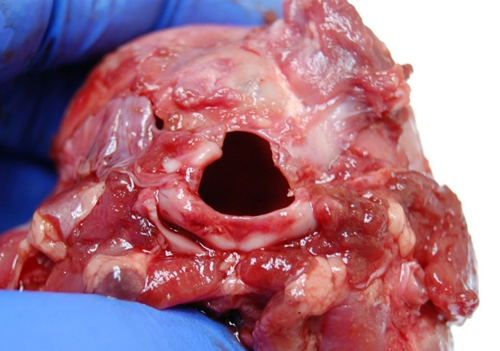
Malformation and ‘keyhole’ shape of the foramen magnum associated with occipital dysplasia was considered the likely cause of sudden death in a nine-week-old Chihuahua puppy
We hope you enjoy reading about our cases of interest. If you would like any help with your own post mortems, please get in touch by phone (01224 711168) or email.
Posted by SRUC Veterinary Services on 31/10/2023