October case highlights
October was a quieter month in the PM room, however submitted lab samples also give us information about disease patterns. Unsurprisingly given the warm, wet back end, and the usual seasonal increase in cases in October, hypomagnesaemia was a notable diagnosis in cattle. Parasitism in lambs continues to be an issue on many farms, reflected both in the egg count submissions and in animals presented for post mortem examination.
Case highlights from the PM room in October include:
- Enterotoxaemia associated with Clostridium perfringens type D infection
- Mesenteric torsion in a dog
- Acute Streptococcus suis Type 1 septicaemia in piglets
Enterotoxaemia associated with Clostridium perfringens type D infection was reported as the cause of death in a seven-week-old suckler calf which died suddenly. The well-grown calf was kept on pasture in a group with 14 cows with calves at foot and was seen playing with the other calves the previous day. The cattle were not vaccinated against clostridial disease. On post mortem examination, dramatic interlobular pulmonary oedema affecting all lobes and a pleural and pericardial effusion were detected (see photo below). The calf had eaten well prior to death, with plenty of grass in the forestomaches and milk clots in the abomasum. Epsilon toxin was detected in the small intestinal contents in the very fresh carcase, which further supported the gross diagnosis of clostridial enterotoxaemia, however microscopic examination of the brain did not detect any significant changes. Histopathological examination of the internal organs was also unremarkable apart from widespread interlobular lung oedema and evidence of coccidiosis in the small and large intestines. It would be unusual for coccidiosis to result in death without prior dysentery or gross lesions in the intestines. In that respect, peracute enterotoxaemia was reported to be strongly suspected as the cause of death, despite the criteria for an official VIDA diagnosis could not been fulfilled. A possible explanation is that epsilon toxin caused severe endothelial damage in the lungs of the calf which cause it to die before brain lesions had an opportunity to manifest. Absence of brain lesions has been reported in other cases of suspect peracute enterotoxaemia.
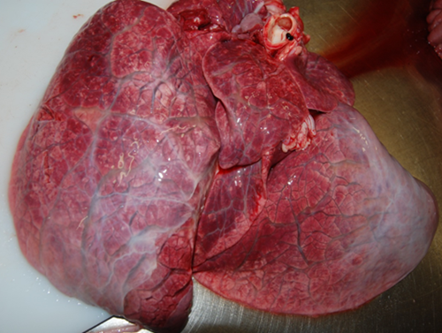
Extensive interlobular oedema affecting all lung lobes
Mesenteric torsion in a dog. A ‘yard dog’, originally from Greece, died suddenly with no history of veterinary treatment since importation. The dog displayed no clinical signs prior to being found dead. On external examination, there was fresh blood around the anus. Internal examination revealed marked congestion of the mesentery and serosa of the small intestine, with copious dark blood stained content within both the small and large intestine, associated with a 180 degree torsion of the mesenteric root. Mesenteric torsion in dogs is rare, and most often seen in larger breeds, with an over-representation of the German Shepherd breed. Intestinal volvulus can be associated with inflammatory bowel disease, intestinal neoplasia, gastrointestinal foreign bodies, recent gastrointestinal surgery, closed abdominal trauma, concurrent gastric dilatation volvulus and boisterous exercise. No concurrent conditions were noted on gross exam, and there was no history consistent with inflammatory bowel disease, therefore boisterous activity was thought to be the most likely predisposing factor in this case. The history of importation from Greece raised the possibility that this dog may have been a subclinical carrier of the zoonotic pathogen Brucella canis, which should be considered if post mortem examinations are carried out in practice.
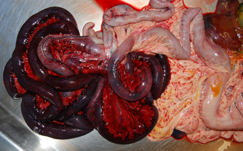
Marked congestion of the mesentery and small intestinal mucosa from the mid-duodenum to the ileocaecal junction, associated with a mesenteric torsion. The affected intestine was thickened and oedematous with darkly blood stained contents.
Acute Streptococcus suis Type 1 septicaemia was diagnosed as the cause of death in two pre-weaned 28-day-old piglets. The piglets derived from a litter of 18, where clinical signs of lethargy, ataxia and cyanosis was followed by death in eight of the piglets. Relatively few gross and histopathological changes were detected apart from suppurative synovitis in several joints of one of the submitted pigs. Streptococcus suis Type 1 was isolated from one of the affected joints, and from the lung, liver, pericardial fluid and brain in the other piglet, which confirmed the diagnosis.
Streptococcus suis is capable of causing a range of disease, including septicaemia (sudden death), meningitis, pneumonia, polyarthritis, and endocarditis and is most commonly seen in five to ten week old piglets. While 29 serotypes have been identified, serotypes 1, 2, 7 and 14 are those most commonly isolated from clinical cases. S. suis outbreaks can be associated with concomitant viral infections, such as with porcine reproductive and respiratory syndrome virus (PRRSV), porcine circovirus 2 (PCV-2) or swine influenza virus (SIV) and also bacterial pathogens such as Bordetella bronchiseptica and Haemophilus (Glaesserella) parasuis. Husbandry factors which predispose to disease outbreaks include changes in husbandry such as moving, mixing or over-crowding and poor ventilation. Control strategies have historically relied on targeted in-feed medication before periods of heightened risk. More recently the promotion of responsible antimicrobial use has encouraged wider deployment of autogenous vaccines.
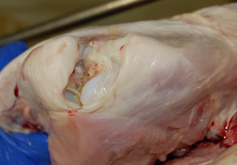
Suppurative synovitis in piglet with Streptococcus suis Type 1 septicaemia.
Posted by SRUC Veterinary Services on 01/12/2023