December case highlights
Bacterial pneumonia dominated the December cattle diagnoses, although two high morbidity pneumonia outbreaks associated with IBR were also investigated. Parasitism remains the most common diagnosis in overwintered growing lambs, as pasture larval populations appear to have survived well into the late Autumn, although the usual overwintering disease such as ruminal acidosis and pasteurella pneumonia also feature in our December diagnoses.
Cases of note from December include:
- Kale toxicity in overwintered lambs
- Salmonella Dublin in a suckler herd
- Enteric yersiniosis in a goat
- Syndactyly with arthrogryposis in a newborn calf
Intravascular haemolysis due to suspect kale toxicity was reported as the cause of death in a 7-month-old ram lamb. Two ram lambs from a group kept outside and fed grass, kale, adlib hay and 100 grams of barley/head had shown clinical signs of ataxia and dyspnoea hours prior to death. Gross findings of jaundice, scarce and watery blood, haemoglobinuria and “gun metal” coloured renal cortex were consistent with intravascular haemolysis. Initial tissue analysis detected unremarkable liver and kidney copper results, which ruled out an interim diagnosis of copper poisoning, the most common cause of haemolysis in sheep. Kale poisoning was therefore suspected based on the clinical history, as consumption of brassicas is a recognised cause of haemolysis in ruminants, when S-methylcysteine sulfoxide (SMCO) is converted to dimethyl disulfide in the rumen. Severity of the disease is proportional to the SMCO content of the crop and when present in small amounts, the toxin results in poor growth rates. Due to the wet weather, more forage crops are being utilised by lambs rather than cattle, and consideration should be given to strip grazing to limit daily intakes and encourage intakes of forage. Parasitic bronchitis, PGE, hyposelenosis and pine was also diagnosed as non-fatal concurrent conditions in the lamb.
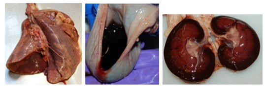
Jaunice, haemoglobinuria and ‘gun metal’-coloured kidneys in a lamb with kale poisoning
Salmonella Dublin septicaemia was diagnosed in a 7-and 15-day-old calf from a suckler herd with split Spring and Autumn calving with bought in replacements. The client reported that all calves born outside from September onwards were not thriving and had various clinical signs including salivation, stiff walking, scour and pyrexia. One cow had similar signs to the calves, but seemed to improve with antibiotic treatment. Gross findings in the two submitted calves were consistent with jaundice, haemorrhage and fibrin exudation and general organomegaly. Isolation of Salmonella Dublin from systemic sites and faeces confirmed the diagnosis. Hypogammaglobulinemia was detected in the youngest calf which is likely to have predisposed to fatal infection. A farm visit was undertaken by SRUC to look at management options for control of S. Dublin in the suckler herd. Salmonellosis is relatively rare in suckler herds and can be a diagnostic challenge due to vague clinical signs (often respiratory disease is the main presenting sign) and difficulty in diagnoses. Post-mortem examination is critical as culture of viscera is the most reliable method of diagnosis (faecal detection is not consistent, particularly where the infection is systemic rather than only enteric). The major risk of entry of S. Dublin into a suckler herd is from added animals, with animals from the dairy sector being a particularly high risk.
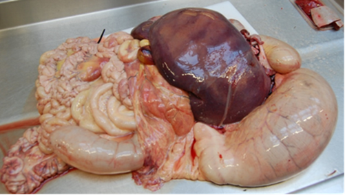
Calf 1: Jaundice and hepatomegaly associated with Salmonella Dublin infection
Calf 2: Splenomegaly with overlying fibrinous exudation associated with Salmonella Dublin infection
Enteric yersiniosis in a goat. A herd of breeding goats experienced the loss of several goats over a one month period, all presenting with acute onset scour and death. Three of the affected animals were yearlings, while the presented animal was an 8 month old kid with a more prolonged disease course, failing to respond to fluids, amoxicillin-clavulanate and steroid treatment. Significant gross findings were confined to an approximately 100cm section of distal jejunum (see pictures) which had marked mural oedema, thickened corrugated mucosa with adherent exudate. The associated lymph nodes were markedly enlarged with 1-4mm diameter white foci mostly on cut surface but visible on the surface of the most proximally affected node. There was a small length of jejunum distal to this which was congested with occasional ulceration. There was ulceration of the ileocaecal junction and the caecum. Yersinia pseudotuberculosis was isolated on bacterial culture from the affected jejunal wall and the lymph node and histopathological examination was consistent with yersiniosis. The client was reminded of the zoonotic potential of Y. pseudotuberculosis and review of shed hygiene and management of risk factors such as stress, poor nutrition, overcrowding and concurrent disease such as parasites, was recommended.
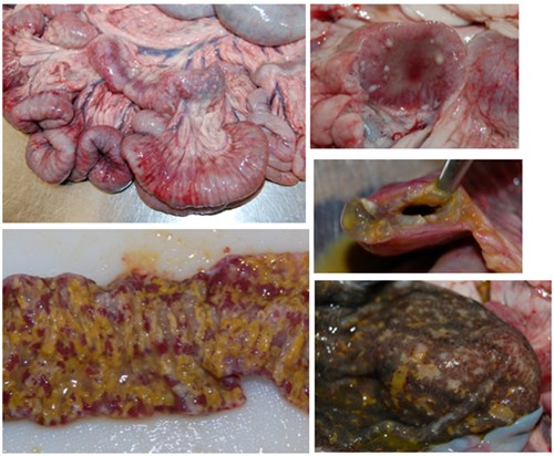
Top left: Section of markedly thickened jejunum with enlarged lymph nodes. Bottom left: washed surface of the affected jejunum. Top right: Cut section of the associated mesenteric lymph node. Middle right: Extent of the mural oedema. Bottom right: Superficial ulceration of the mucosal surface of the caecum.
Syndactyly and arthrogryposis associated with cerebellar dysplasia. A large herd of predominantly limousin cross suckler cows were kept in stable management groups and put to the same bull annually. Four calves have been born this year, two in Spring and two of the current back end calvers. all with the same sire, and presented with similarly deformed legs and single digits (syndactyly) affecting all 4 feet. The bull was 8 years old and had bred with the same groups of cows (approx. 55 cows per year) for 4 years previously without any issues. The gross examination was as described in the legend below. Cerebellar dysplasia was found on neuropathological examination and likely contributed to the development of arthrogryposis. Pestivirus PCR was negative.
Syndactyly in cattle, also known as 'mulefoot', is inherited as an autosomal recessive trait with variable penetrance in different cattle breeds. Responsible genes have been identified in the Holstein Friesian and Simmental cattle, however it has also been reported in Angus, Chianina, Hereford, German Red Pied, Indian Hariana, and Japanese native cattle. The presence of the condition in both the spring and autumn calvers, and restricted to the calves of one bull with the cows despite a shared winter ration makes a heritable cause very likely here. The factors that lead to incomplete penetrance of the condition, and particularly why so many clinical cases have occurred this year and not in previous years, are unknown however. 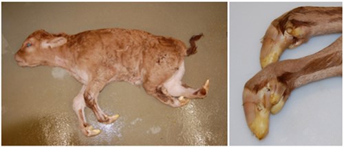
The mandible and maxilla appeared proportionally moderately shortened. There was syndactyly of all four feet. The forelimbs were slightly short with mild arthrogryposis leading to flexion of the carpi and digits. With the exception of the femurs, the bones hindlimbs were markedly shortened, especially the distal limbs which were also medially rotated, and somewhat fused in a flexed position. The accessory claws were prominent/overdeveloped in all feet. There was a small vestigial remnant of the missing claw with the exception of the left hind where the remnant was larger and was fused to the side of the formed claw.
Posted by SRUC Veterinary Services on 29/02/2024