August case highlights
August was another steady month in the Aberdeen PM room, with the predominant submission being thin or dying lambs with parasitic gastroenteritis. The wet summer certainly helped parasites complete their life cycle efficiently and heavy pasture larval burdens caught a number of farmers out, especially those relying on fixed time dosing. The worm problems weren’t confined to sheep, and vet services are seeing increased cases of parasitism in cattle, both in young stock and adult animals.
Other interesting cases from the PM room in August include:
- Pneumonia in dairy-beef cross rearing calves
- Inflammatory pericarditis leading to pericardial effusion in a sheep
- Suspect ectodermal dysplasia in an aborted calf
Bovine respiratory disease complex in dairy-beef calves. Three submissions from different holdings experiencing on-going losses due to bovine respiratory disease (BRD) were examined in Aberdeen. These were all purchased dairy-cross calves aged between three and nine months being reared in housed groups for finishing. Acute and chronic, non-responsive respiratory disease was a feature in these herds. Bacterial culture was generally unrewarding, attributed to ante-mortem antibiotic use. Bovine Respiratory Disease Multiplex-Tandem PCR detected significant concentrations of Mycoplasma bovis, Histophilus somni, Pasteurella multocida and Mannheimia haemolytica DNA in all of the presented calves, with Respiratory Syncytial Virus RNA also detected in one calf. Two of the affected farms were vaccinating calves against M. bovis on arrival and on one farm vaccination for H. somni and M. haemolytica occurred concurrently. The third farm vaccinated against viral pneumonia only. These cases show the challenges of calf dairy-beef calf rearing, especially with respect to managing respiratory disease. They reinforce that while vaccination and early, effective treatment protocols have a role in controlling respiratory disease, optimal neonatal calf management on the holding of origin and management of stressors during rearing form an essential part in the prevention and management of respiratory disease.
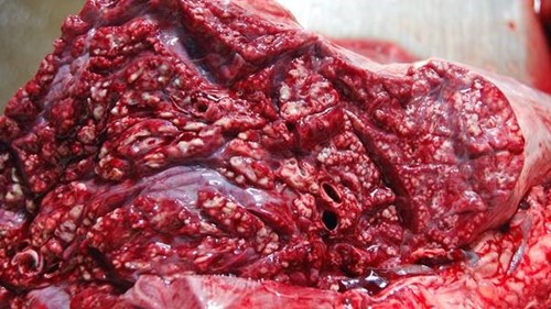
Microabscessation of consolidated lung tissue associated with M. bovis, H. somni, P. multocida and M. haemolytica
Fibrinopurulent epicarditis / pericarditis resulting in haemorrhagic pericardial effusion in a four-year-old Valais cross ewe. The ewe was from a 14 ewe hobby flock and had a two month history of lethargy, submandibular oedema and pale mucous membranes, with acute deterioration prior to death. On gross examination the carcase was pale with depleted adipose stores. The pericardium was markedly distended with 1.3 litres of blood stained fluid. There was an irregular five centimetre by three centimetre granular lesion of the right auricle with adherent stringy blood clot and similar adherent granulomatous lesions present diffusely over the epicardial surface and those of the great vessels. There was a ‘nutmeg’ pattern on cut surface of the liver and approximately six to eight litres of clear effusion in the peritoneal cavity and a mild pleural effusion, consistent with congestive failure associated with cardiac tamponade. On the epicardial surface of the auricle, and pericardium, there were multifocal areas of mesothelial proliferation, fibrin deposits and neutrophils seen histopathologically indicating a fibrinopurulent peri- and epicarditis with mesothelial hyperplasia. Examination of systemic tissues revealed no evidence of a systemic inflammatory or infectious process. The cardiac lesions were thought most likely to have originated from a sporadic infectious pericarditis.
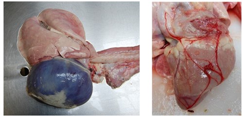
Left: distended pericardium. Right: Granulomatous lesion on the right auricle with adherent clot with diffuse tiny granulomatous lesions over the epicardial surface.
Suspect genetodermatosis/ectodermal dysplasia born to an imported heifer. A group of imported in-calf heifers were introduced to a large suckler cow herd and formed part of 28 cow group, where two animals had aborted. The calf from the second heifer to abort was examined, with marked alopecia sparing the peripheral skin the most notable finding. Gross and histopathological examination of the thyroid was unremarkable and fungal culture was negative. Histopathological examination of the tissues including skin sections was suggestive of ectodermal dysplasia. These genetic conditions affect the development and/or homeostasis of two or more ectodermal derivatives, including hair, teeth, nails, and eccrine glands. In one report of calves with ectodermal dysplasia there was also an absence of nasolabial, intranasal and tracheobronchial mucosal glands, predisposing these calves to respiratory infections which led to the death of most calves within ten days of birth. There are no reports of this condition leading to abortion, however a novel genetic defect which incorporates an ectodermal dysplasia and affects foetal viability could not be excluded. SRUC Veterinary Services expressed interest in further investigation should any further cases occur on farm.
Reference: O’Toole, D., Häfliger, I.M., Leuthard, F., Schumaker, B., Steadman, L., Murphy, B., Drögemüller, C. and Leeb, T., 2021. X-linked hypohidrotic ectodermal dysplasia in crossbred beef cattle due to a large deletion in EDA. Animals, 11(3), p.657.
Posted by SRUC Veterinary Services on 04/10/2023